A World Under the Microscope: Nikon Reveals Winners of Small World 2022 Photomicrography Awards

A World Under the Microscope: Nikon Reveals Winners of Small World 2022 Photomicrography Awards
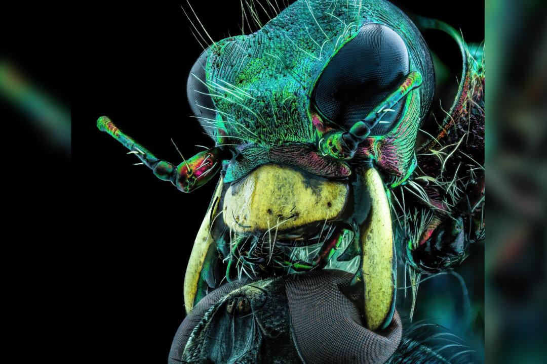
Courtesy of Murat Öztürk and Nikon Small World
Updated:

Three top winners of Nikon’s Small World 2022 Photomicrography Competition alongside dozens of notable entries were recently released, showcasing a world of limitless detail under powerful magnifying lenses.

The top pictures were picked from some 1,300 entries by scientists and artists, amateurs and professionals, representing 72 different countries. The dazzling array of images proves there is beauty and art under the microscope, and more sublimity in the fine details than we could have imagined.

A panel of experts in the fields of art and biology awarded the top prize in the 48th annual competition to Grigorii Timin, under Dr. Michel Milinkovitch at the University of Geneva, Switzerland, for his photo of a tiny reptilian foot, belonging to a Madagascar giant day gecko, still in the early stages of development.

Timin used a high-resolution confocal microscope and the complex process of image-stitching to tie hundreds of pictures together for his final submission, according to a Nikon news release. Even assembling the collage was a challenge, Timin said.
“This embryonic hand is about 3mm in length, which is a huge sample for high-resolution microscopy,” he said. “The scan consists of 300 tiles, each containing about 250 optical sections, resulting in more than two days of acquisition and approximately 200GB of data.”

1st Place

Embryonic hand of a Madagascar giant day gecko (<span class="font6">phelsuma grandis</span><span class="font5">), taken by Grigorii Timin and Dr. Michel Milinkovitch.</span> (Courtesy of Grigorii Timin and Dr. Michel Milinkovitch and Nikon Small World)
Embryonic hand of a Madagascar giant day gecko (phelsuma grandis), taken by Grigorii Timin and Dr. Michel Milinkovitch. Courtesy of Grigorii Timin and Dr. Michel Milinkovitch and Nikon Small World

Timin’s submission, a sprawling display of glowing ochre set off by cool turquoise, captures nerves, bones, tendons, ligaments, skin, and blood cells of the gecko’s miniscule foot in mind-blowing detail.

“This particular image is beautiful and informative as an overview, and also when you magnify it in a certain region, shedding light on how the structures are organized on a cellular level,” Timin said.

Second place went to Dr. Caleb Dawson of Melbourne, Australia, for his photo of breast tissue, detailing minute contractile myoepithelial cells wrapped around milk-producing alveoli. Dawson also used a confocal microscope. He then spent a week staining myoepithelial cells with multiple rounds of fluorescent dye to capture this wonderous image.

2nd Place

Breast tissue showing contractile myoepithelial cells wrapped around milk-producing alveoli, taken by Dr. Caleb Dawson. (Courtesy of Caleb Dawson and Nikon Small World)
Breast tissue showing contractile myoepithelial cells wrapped around milk-producing alveoli, taken by Dr. Caleb Dawson. Courtesy of Caleb Dawson and Nikon Small World

3rd Place

Blood vessel networks in the intestine of an adult mouse, taken by Satu Paavonsalo and Dr. Sinem Karaman. (Courtesy of Satu Paavonsalo and Dr. Sinem Karaman and Nikon Small World)
Blood vessel networks in the intestine of an adult mouse, taken by Satu Paavonsalo and Dr. Sinem Karaman. Courtesy of Satu Paavonsalo and Dr. Sinem Karaman and Nikon Small World

Third place was handed to another collaboration; Satu Paavonsalo and Dr. Sinem Karaman of the University of Helsinki, Finland, captured a photo of intestinal blood vessel networks in an adult mouse.

There were several notable mentions, including award-winning Lithuanian photographer Eugenijus Kavaliauskas, who portrayed a chilling close-up of a carpenter ant’s head. This photo, honored with a distinction in the competition, quickly went viral on social media, and Twitter user Rebekah McKendry PhD likened the ant to an “image from a horror movie.”
Kavaliauskas told The Washington Post his photo was achieved by magnifying the ant’s face five times under a stereo 10x microscope. He described the image as an example of “God’s designs and the many interesting, beautiful, unknown miracles under people’s feet ... there are no horrors in nature, only lack of knowledge.”

10th Place

A fly under the chin of a tiger beetle, taken by Murat Öztürk. (Courtesy of Murat Öztürk and Nikon Small World)
A fly under the chin of a tiger beetle, taken by Murat Öztürk. Courtesy of Murat Öztürk and Nikon Small World

Another portrait of a teeny insect’s head, that of a tiger beetle, seems ginormous as it fills the frame. Its insignificant mandibles seem formidable as they clamp down on the head of a puny fly. Additionally, the judges picked up-close images of glowing red algae, microscopic crystal, unburned carbon particles being released from a candle wick, and delicate moth eggs. The range of awe-inspiring microscopic subject matter is jaw-dropping.

There are indeed countless unseen worlds under our noses in the most seemingly insignificant places.

4th Place

Long-bodied cellar/daddy long-legs spider (<span class="font6">pholcus phalangioides</span><span class="font5">), taken by Dr. Andrew Posselt.<. (Courtesy of Dr. Andrew Posselt and Nikon Small World)</span>
Long-bodied cellar/daddy long-legs spider (pholcus phalangioides), taken by Dr. Andrew Posselt.<. (Courtesy of Dr. Andrew Posselt and Nikon Small World)

5th Place

Slime mold (l<span class="font6">amproderma</span><span class="font5">), taken by Alison Pollack. (Courtesy of Alison Pollack and Nikon Small World)</span>
Slime mold (lamproderma), taken by Alison Pollack. (Courtesy of Alison Pollack and Nikon Small World)

6th Place

Unburned particles of carbon released when the hydrocarbon chain of candle wax breaks down, taken by Ole Bielfeldt. (Courtesy of Ole Bielfeldt and Nikon Small World)
Unburned particles of carbon released when the hydrocarbon chain of candle wax breaks down, taken by Ole Bielfeldt. Courtesy of Ole Bielfeldt and Nikon Small World

7th Place

Human neurons derived from neural stem cells, taken by Dr. Jianqun Gao and Prof. Glenda Halliday. (Courtesy of Dr. Jianqun Gao and Prof. Glenda Halliday and Nikon Small World)
Human neurons derived from neural stem cells, taken by Dr. Jianqun Gao and Prof. Glenda Halliday. Courtesy of Dr. Jianqun Gao and Prof. Glenda Halliday and Nikon Small World

8th Place

Growing tip of a red algae, taken by Dr. Nathanaël Prunet. (Courtesy of Dr. Nathanaël Prunet and Nikon Small World)
Growing tip of a red algae, taken by Dr. Nathanaël Prunet. Courtesy of Dr. Nathanaël Prunet and Nikon Small World

9th Place

A liquid crystal mixture, taken by Dr. Marek Sutkowski. (Courtesy of Dr. Marek Sutkowski and Nikon Small World)
A liquid crystal mixture, taken by Dr. Marek Sutkowski. Courtesy of Dr. Marek Sutkowski and Nikon Small World

11th Place

Moth eggs, taken by Ye Fei Zhang. (Courtesy of Ye Fei Zhang and Nikon Small World)
Moth eggs, taken by Ye Fei Zhang. Courtesy of Ye Fei Zhang and Nikon Small World

12th Place

Autofluorescence of a single coral polyp, taken by Brett M. Lewis. (Courtesy of Brett M. Lewis and Nikon Small World)
Autofluorescence of a single coral polyp, taken by Brett M. Lewis. Courtesy of Brett M. Lewis and Nikon Small World

13th Place

Agatized dinosaur bone, taken by Randy Fullbright. (Courtesy of Randy Fullbright and Nikon Small World)
Agatized dinosaur bone, taken by Randy Fullbright. Courtesy of Randy Fullbright and Nikon Small World

14th Place

Differentiated cultured mouse myoblasts with lysosomes (cyan/green), nuclei (yellow), and F-actin (magenta), taken by Nadia Efimova. (Courtesy of Nadia Efimova and Nikon Small World)
Differentiated cultured mouse myoblasts with lysosomes (cyan/green), nuclei (yellow), and F-actin (magenta), taken by Nadia Efimova. Courtesy of Nadia Efimova and Nikon Small World

15th Place

Cross sections of normal human colon epithelial crypts, taken by Dr. Ziad El-Zaatari. (Courtesy of Dr. Ziad El-Zaatari and Nikon Small World)
Cross sections of normal human colon epithelial crypts, taken by Dr. Ziad El-Zaatari. Courtesy of Dr. Ziad El-Zaatari and Nikon Small World

16th Place

Longitudinal section through a white asparagus shoot tip, taken by Dr. Olivier Leroux. (Courtesy of Dr. Olivier Leroux and Nikon Small World)
Longitudinal section through a white asparagus shoot tip, taken by Dr. Olivier Leroux. Courtesy of Dr. Olivier Leroux and Nikon Small World

17th Place

Tail fin of a zebrafish larva with peripheral nerves (green) and extracellular matrix (violet), taken by Dr. Daniel Wehner and Julia Kolb. (Courtesy of Dr. Daniel Wehner and Julia Kolb and Nikon Small World)
Tail fin of a zebrafish larva with peripheral nerves (green) and extracellular matrix (violet), taken by Dr. Daniel Wehner and Julia Kolb. Courtesy of Dr. Daniel Wehner and Julia Kolb and Nikon Small World

18th Place

Network of macrophages (white blood cells) of an adult zebrafish intestine, taken by Dr. Julien Resseguier. (Courtesy of Dr. Julien Resseguier and Nikon Small World)
Network of macrophages (white blood cells) of an adult zebrafish intestine, taken by Dr. Julien Resseguier. Courtesy of Dr. Julien Resseguier and Nikon Small World
Bacterial biofilm on a human tongue cell, taken by Dr. Tagide deCarvalho. (Courtesy of Dr. Tagide deCarvalho and Nikon Small World)
Bacterial biofilm on a human tongue cell, taken by Dr. Tagide deCarvalho. Courtesy of Dr. Tagide deCarvalho and Nikon Small World

2oth Place

Human cardiomyocytes (heart cells) derived from induced pluripotent stem cells, taken by Hui Lin and Dr. Kim McBride. (Courtesy of Hui Lin and Dr. Kim McBride and Nikon Small World)
Human cardiomyocytes (heart cells) derived from induced pluripotent stem cells, taken by Hui Lin and Dr. Kim McBride. Courtesy of Hui Lin and Dr. Kim McBride and Nikon Small World
Share your stories with us at [email protected], and continue to get your daily dose of inspiration by signing up for the Inspired newsletter at TheEpochTimes.com/newsletter