Understanding Veteran TBI: Hormone Imbalances Following Traumatic Brain Injury

Understanding Veteran TBI: Hormone Imbalances Following Traumatic Brain Injury
A self portrait of Jonathan David Chandler. One half of the image shows him in his military uniform and was taken while receiving treatment for TBI suffered while serving in Afghanistan. The second half is an MRI scan. DoD photo by Petty Officer 2nd Class Jonathan David Chandler
Geoffrey P. Dardia
Battlefields Staff
Updated:
0:00
Commentary

It has been known for over 80 years that following a head injury there is dysfunction in the hypothalamic-pituitary-adrenal (HPA) axis. According to the Centers for Disease Control (CDC), 80–85 percent of traumatic brain injury (TBI) sufferers are classified as mild traumatic brain injuries (MTBI). Approximately 80–85 percent of MTBI patients recover from their injuries, while approximately 15–20 percent continue to experience persistent symptoms. Fifty percent of those TBI patients end up with some type of hormonal imbalance in the HPA axis. This brain injury-related condition is commonly referred to as neuroendocrine dysfunction (NED).
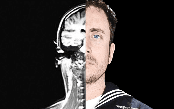
The United States military has been plagued by suicides, TBIs, and post-traumatic stress disorder (PTSD) for the past 20 years. The human toll and financial costs associated with these conditions are crippling the Department of Defense (DoD). As of 2017, the DoD still has not implemented mandatory NED testing following a TBI. Countless veterans and active duty service members could have suffered traumatic brain injuries while serving their country, without ever being properly screened, diagnosed, and treated for TBI. Many veterans could also have been discharged for conditions related to their brain injuries, and more than likely could have continued serving if they had been properly screened, diagnosed, and treated for their injuries. Many service members are also chronically medicated for the symptoms associated with TBI with no resolution of their underlying health issues.

Most of the symptoms associated with TBI and PTSD can be traced back to hormonal imbalances in the HPA axis, and to dysfunction in the autonomic nervous system (ANS). NED and ANS dysfunction can both be screened to show if a person is at risk for PTSD, suicide, or if they have suffered a brain injury in the past. Early and accurate detection of TBI, utilizing advanced brain scanning techniques like diffusion tensor imaging (DTI), quantitative single photon emitting computed tomography (qSPECT), and advanced laboratory testing of biomarkers is critical for timely and optimal patient recovery. The hypothalamic-pituitary-adrenal axis is a crucial system that regulates nearly every system in the body. Before you can fully understand the magnitude of NED, you must understand the function of each gland in the axis and the roles they play that directly affect your life.

The pituitary gland, which is known as the master gland, produces hormones that regulate other glands in the body, such as the thyroid gland, adrenal glands, testes, and ovaries. The pituitary gland is the size of a pea and it is responsible for regulating almost every system in the body via the hypothalamus. The anterior lobe of the pituitary gland produces the following hormones: luteinizing hormone (LH), follicle stimulating hormone (FSH), thyroid stimulating hormone (TSH), prolactin (PRL), adrenocorticotropic hormone (ACTH), growth hormone (GH), and lastly, beta-endorphins. The posterior lobe of the pituitary gland stores anti-diuretic hormone (ADH) and oxytocin (OT). These hormones regulate functions such as sexual reproduction, arousal, stress response, pain response, muscle development, immune function, body composition, cognitive function, metabolism, body temperature, behavior, and most importantly, homeostasis. One of the most common and unknown conditions associated with TBI is growth hormone deficiency (GHD). Imbalances of pituitary hormones can lead to infertility, hypogonadism (low testosterone), adrenal insufficiency, hypothyroidism, type II diabetes, dyslipidemia (abnormal cholesterol in the blood) and hypertension (high blood pressure).

Although the pituitary gland is considered the master gland, it is controlled by the hypothalamus. The hypothalamus is the size of a walnut. Located at the base of the brain, it is part of the limbic system, which plays important roles in behavior, mood, memory, motivation, and situational awareness.

The hypothalamus controls the ANS and the endocrine system via the pituitary gland. The hypothalamus gland produces releasing hormones, which signal the pituitary gland to produce hormones as needed for the body to react to the environment.

The adrenal glands produce three main hormones: cortisol, aldosterone, and adrenaline. The adrenal glands are located on top of the kidneys and produce the hormones aldosterone, cortisol, and adrenaline. Cortisol, also known as the “stress hormone” is responsible for many critical functions throughout the body, including metabolism, glucogenesis (the formation of glucose through the breakdown of glycogen), immune function, inflammation, blood pressure, and most importantly, the nervous system. Cortisol levels are directly influenced by stress in our environment. Cortisol production takes priority in the autonomic nervous system over sex hormones like testosterone when in a sympathetic response state. The steroid hormone pathway, in a sympathetic state, switches to the production of glucocorticoids like cortisol to keep you alert and alive for your “fight or flight” response. Elevated cortisol levels are associated with being hypervigilant, while low cortisol levels are associated with depression, apathy, or feeling numb. When cortisol is produced, testosterone production decreases, this is known as the “pregnenolone steal.”

Aldosterone is a mineralocorticoid hormone produced in the adrenal glands. Aldosterone is responsible for regulating fluid, sodium, and potassium levels in the body. Aldosterone imbalance can directly affect blood pressure, electrolyte balance, and water retention in the body, causing cramps, bloating, or swelling in the extremities.

Adrenaline, also known as epinephrine, activates the sympathetic response (fight or flight) in the ANS and is responsible for the following functions: it increases strength, dilates the pupils, increases heart rate, increases respiratory rate, opens airways, lowers body temperature, shunts blood to your vital organs, numbs arousal, dulls pain, and heightens awareness. When the body is in a constant state of stress, adrenaline and cortisol remain elevated, which in turn suppresses metabolism, immune function, sexual arousal, sex organ function, increases inflammation, increases blood glucose levels, decreases digestion and absorption, increases blood pressure, decreases cognitive functioning, hinders short term memory and causes sleep disturbances.

The DoD spends nearly $7 billion on prescription drugs annually for conditions that can be directly attributed to TBI or stress related neuroendocrine related imbalances. The collateral damage of unnecessary or improper use of prescription drugs is taking its toll on the force in the form of suicides, adverse drug related deaths, and the exorbitant DoD budget for prescription drugs. Most neuroendocrine imbalances can be corrected by lifestyle and environmental adjustments or managed with precision medicine. Treating the symptoms of hormone imbalances has proven costly, deadly, and ineffective, as 15–25 percent of TBI patients see no resolution to their conditions. Out of frustration and desperations, service members and veterans often resort to the use of dangerous and deadly street drugs, prescription drugs, and alcohol as a form of self-medication.

References

Agha A., Rogers B., Mylotte D., Taleb F., Tormey W., Phillips J., Thompson C.J. (2004, May). Neuroendocrine dysfunction in the acute phase of traumatic brain injury.
Klose M., Juul A., Struck J., Morgenthaler N.G., Kosteljanetz M., Feldt-Rasmussen U. (2007, October). Acute and long-term pituitary insufficiency in traumatic brain injury: a prospective single-centre study.
Tölli A., Borg J., Bellander B.M., Johansson F., Höybye C. (2017, February). Pituitary function within the first year after traumatic brain injury or subarachnoid haemorrhage.
The appearance of U.S. Department of Defense (DoD) visual information does not imply or constitute DoD endorsement.
This article first appeared in The Havok Journal.
Master Sergeant Geoffrey P. Dardia is a career Special Forces soldier currently serving as the Operations Sergeant for 3rd Special Forces Group’s Human Performance and Wellness Program. MSG Dardia is the founder and action officer for 3rd Special Forces Group’s META Retirement and Transition Program. He served in both the Central Command (CENTCOM) and U.S. Africa Command (AFRICOM) areas of operation as a Special Forces advisor, and as a liaison at the U.S. Embassy in Burkina Faso. He is also a Veteran’s Affairs Certified Recovery Care Coordinator (RCC) and served as the Ft. Bragg Wounded Warrior Battalion Liaison for United States Special Operations Command (USSOCOM). MSG Dardia cowrote and published his first scientific paper in 2019 in the Alternate Therapies in Health and Medicine (ATHM) Journal titled “Neurotoxicity Associated with Traumatic Brain Injury, Blast, Chemical, Heavy Metal and Quinoline Drug Exposure.”
Related Topics