Heparan Sulfate and the Brain Ventricles
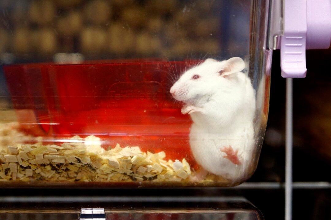
The fact that a manipulation so specific to heparan sulfate in the brain is enough to induce autism in mice suggests that brain deficiencies in heparan sulfate may be a key central pathology in human autism. Indeed, many genetic mutations linked to autism involve enzymes associated with the synthesis of the so–called extracellular matrix [10]. This is the non–cellular component of tissues and organs, which not only provides physical scaffolding but also initiates and orchestrates many biomechanical and biochemical cues that govern cell physiological responses to environmental stimulants [11]. A number of the mutations linked to human autism occur in a set of genes that are called “glycogenes,” which encode the proteins and lipids that are bound to heparan sulfate in the matrix, forming “heparan sulfate proteoglycans” (HSPGs), or enzymes involved in “glycosylation” – the binding of heparan sulfate and similar complex sugar chain molecules to these proteins and lipids [10].The ventricles of the brain are a network of cavities in the middle of the brain that are filled with cerebrospinal fluid. Heparan sulfate (HS) is prominent in the ventricles, found within structures called “fractones,” making up the stem cell niche that initiates neurogenesis [12]. Under the guidance of HSPGs within these specialized extracellular matrix zones, stem cells proliferate and differentiate into specialized cells and migrate into the brain to replace damaged neurons. Studies on mice have shown that disruption of an enzyme that is essential for synthesis of HS in the early developmental stages of mouse embryos results in severe disruption of brain development [13].
BTBR Mice: Gut Issues
A seminal study on these BTBR mice revealed specific disruptions in the gut that were hypothesized to lead to the neurological effects through interactions along the gut–brain axis [18]. The most glaring disorder observed was a disruption in the synthesis of bile acids in the liver and of their further modification by gut bacteria. Normally, the liver synthesizes bile acids from cholesterol and conjugates them with either taurine or glycine before shipping them out to the gut or buffering them in the gall bladder. It is the responsibility of specific species of gut bacteria, mainly Bifidobacteria, to deconjugate the conjugated bile acids, freeing up the taurine or glycine molecule for further metabolism. This is a necessary step before the bile acids can be further modified by other gut bacteria, notably the species Blautia, into secondary bile acids. Thus there are many different variants of the bile acids, and the distinct forms have differing signaling effects influencing peristalsis and gut barrier integrity.Did Glyphosate Cause Autism in BTBR Mice?
It is easy to argue that these abnormalities could be due in part to glyphosate exposure. These mice are the progeny of multiple generations of inbred lab mice that were almost certainly fed a steady diet of glyphosate in their mouse feed manufactured from genetically modified Roundup–Ready corn and soy crops. A reduced supply of bile acids in each generation, and direct toxicity of glyphosate to certain species of bacteria, would alter the microbial distribution over time. Thus, the gut microbes that were passed on from generation to generation could maintain a pathological distribution influenced by glyphosate acting as an antibiotic and enzyme disruptor [19].Bile acid synthesis depends crucially on cytochrome P450 (CYP) enzymes in the liver. Glyphosate has been shown to severely reduce CYP enzyme expression in the rat liver [19, 20]. A study on poultry microbiota showed that Bifidobacteria were especially highly sensitive to glyphosate, compared to all other species examined [21]. It is logical that Bifidobacteria would suffer from glyphosate exposure due to their role in deconjugating bile acids, because glyphosate can be expected to substitute for glycine during the conjugation step, due to the fact that it is an amino acid analogue of glycine [22, 23]. Bifidobacteria would be tasked with deconjugating glyphosate from bile acids, and then would be directly exposed to the liberated glyphosate molecule.
Studies on Mice Exposed to Glyphosate
Exposure of male mice to glyphosate–based herbicides during the juvenile and adult period led to a marked reduction in serotonin levels in several nuclei in the brainstem [26]. This was associated with weight loss, decreased locomotor activity, and an increase in anxiety and depression–like behavior. Serotonin, whether produced in the brain or the gut, is sulfated in transit, and melatonin, which is derived from serotonin, is also sulfated. We argued in a paper published in 2015 that glyphosate could collaborate with aluminum to induce both gut dysbiosis and disruption of pineal gland function in the brain [2]. The pineal gland produces sulfated melatonin and distributes it into the cerebrospinal fluid of the ventricles during sleep. We proposed that an important role for melatonin is to deliver sulfate to neurons to boost the sulfate supplies in the HSPGs. Heparan sulfate plays a significant role in the clearance of cellular debris, which is an important aspect of sleep. And sleep disturbance is a common feature of autism [27]. So this is getting close to closing the gap between the heparan sulfate deficiency observed in the brains of BTBR mice and their gastrointestinal disturbances.Taurine: Miracle Molecule?
Even before I knew the word glyphosate, I published an article, together with other colleagues, titled: “Is Encephalopathy a Mechanism to Renew Sulfate in Autism?” [28]. In this paper, we discussed the crucial role of heparan sulfate in the brain and a potential link to autism. We proposed that taurine plays a central role in restoring sulfate supplies to the brain under stressful conditions. Curiously, human cells are unable to metabolize taurine, but dietary taurine can get converted to sulfate by gut microbes. The brain, heart and liver all store large amounts of taurine, and this taurine is released into circulation during encephalopathy (brain swelling) or during a heart attack. This taurine is then taken up by the liver and conjugated to bile acids. The taurine, received by the deconjugating gut microbes, can then be oxidized to sulfate, to boost supplies in the blood. I suspect, although at this time this is only speculation, that the bile acids serve a crucial role in facilitating the reaction that releases the sulfonate moiety from taurine, perhaps by anchoring the taurine molecule in the bacterial membrane. Further oxidation by sulfite oxidase yields sulfate. Glyphosate’s damaging effects on Bifidobacteria would interfere with the production of sulfate from taurine by gut microbes, due to impairment in the ability to detach taurine from the bile acids.Clostridia Overgrowth and Vaccine–Induced Autism
A very different mouse model of autism involves exposure of a pregnant mouse dam to virus–like particles during gestation. Two publications describing one such experiment have gained considerable attention from the media, particularly because they demonstrated a link between a particular profile of gut microbial colonization in the dam and a susceptibility to autism in the pups [7, 8]. The pups not only exhibited classic autistic behavior, but also had “patches of disorganized cortical cytoarchitecture” within a specific region in the somatosensory cortex of their brains, showing disrupted brain development architecturally.The authors noted that the autistic profile only arose if the dam had an over–representation of a specific filamentous Clostridia strain in the gut, which in turn led to expression of a “Th17” type immune response by the dam’s immune system. A communication between the gut and brain led, remarkably, to a signaling cascade that had a direct impact on the developing fetuses. The virus–like particles, called “polyinosinic:polycytidylic acid” (poly(I:C)) were injected into the brain of the dam on embryonic day 12.5. These particles are not a life form, but they fool the brain’s immune system into believing that there has been a viral invasion in the brain, and it is the immune response itself, not the viral infection, that induces the overactive response adversely affecting brain development in the offspring. And, what is even more surprising is that the defects develop in the mouse pups only if there is a particular distribution of gut microbes favoring the filamentous Clostridia species.
An earlier study using this same mouse model of injecting a pregnant dam with poly(I:C) links Clostridia overgrowth to the release of certain specific toxins, and, remarkably, links these toxins directly to autism [17]. Several species of Clostridia produce toxic phenolic metabolites such as 4–ethylphenyl sulfate (4EPS) and p–cresol sulfate. The offspring of exposed mouse dams displayed a striking 45–fold increase in serum levels of 4EPS, as well as elevated levels of p–cresol sulfate. This was associated with elevated levels of inflammatory factors in the maternal blood, placenta and aminiotic fluid. Notably, a 3–week treatment of young healthy mice with 4EPS potassium salts was enough to induce autistic symptoms in these mice. Furthermore, probiotic treatment with the species Bacteroides fragilis ameliorates autistic symptoms in the offspring of poly(I:C) exposed dams.
Human Studies are Consistent with the Mouse Studies
A recent study by William Shaw involved a set of triplets, two boys and a girl [31]. Both boys were diagnosed with autism and the girl had a seizure disorder. All three children were found to have high levels of glyphosate in their urine. They also had overrepresentation of Clostridia species in the gut, which were suggested to contribute to the disease process through their release of toxic phenolic metabolites. Another study from 2017 on the gut microbiome of autistic children with inflammatory bowel disease compared to normal controls showed reduced Blautia species (impaired bile acid metabolism) and increases in several species of Clostridia that were linked to reduced tryptophan levels and impaired serotonin homoestasis, along with overexpression of Th17, all consistent with the various mouse model studies [32].Conclusion
In summary, a disrupted gut microbiome (which can be caused by glyphosate) leads to a leaky gut barrier, a leaky brain barrier and a leaky placental barrier. This allows toxic substances such as aluminum, phenolic compounds and glyphosate, as well as live viruses and endotoxins from vaccines, to invade the brain, and, by breaching the placental barrier, expose the fetus to harm. An overzealous immune reaction to these insults disrupts neuronal development and causes autistic–like behaviors in the mouse pups and in children whose mothers have been similarly exposed.The BTBR mice became autistic after many generations of inbreeding during glyphosate exposure in the lab. It would be very interesting to find out what would happen if a group of BTBR mice were provided a nutrient–dense organic diet and clean water, and allowed to reproduce through multiple generations with this healthy diet. Would the descendants eventually lose their autism diagnosis? If they did, it would tell us a great deal about the importance of an organic diet to human health, and would greatly strengthen the idea that glyphosate is a causative factor in autism
References
[1] Swanson N, Leu A, Abrahamson J, Wallet B. Genetically engineered crops, glyphosate and the deterioration of health in the United States of America. Journal of Organic Systems 2014; 9: 6–37.[2] Seneff S, Swanson N, Li C. Aluminum and Glyphosate Can Synergistically Induce Pineal Gland Pathology: Connection to Gut Dysbiosis and Neurological Disease. Agricultural Sciences 2015; 6: 42–70.
[3] Beecham JE, Seneff S. Is there a link between autism and glyphosate–formulated herbicides? Journal of Autism 2016; 3:1.
[4] Beecham JE, Seneff S. The Possible Link between Autism and Glyphosate Acting as Glycine Mimetic – A Review of Evidence from the Literature with Analysis. J Mol Genet Med 2015; 9:4
[5] McFarlane HG, Kusek GK, Yang M, Phoenix JL, Bolivar VJ, Crawley JN. Autism–like behavioral phenotypes in BTBR T+tf/J mice. Genes Brain Behav. 2008;7(2):152–63. Epub 2007 Jun 7.
[6] Scattoni ML, Ricceri L, Crawley JN. Unusual repertoire of vocalizations in adult BTBR T+tf/J mice during three types of social encounters. Genes Brain Behav 2011; 10:44–56.
[7] Kim S, Kim H, Yim YS, Ha S, Atarashi K, Tan TG, Longman RS, Honda K, Littman DR,, Choi GB, Huh JR. Maternal gut bacteria promote neurodevelopmental abnormalities in mouse offspring. Nature 2017;549: 528–532.
[8] Yim YS, Park A, Berrios J, Lafourcade M, Pascual LM, Soares N, Kim JY, Kim S, Kim H, WSaisman A, Littman DR, Wickersham IR, Harnett MT, Huh JR, Choi GB. Reversing behavioural abnormalities in mice exposed to maternal inflammation. Nature 2017;549: 482–487.
[9] Irie F, Badie–Mahdavi H, Yamaguchi Y. Autism–like socio–communicative deficits and stereotypies in mice lacking heparan sulfate. Proc Natl Acad Sci U S A. 2012 Mar 27; 109(13): 5052–5056.
[10] Dwyer CA, Esko JD. Glycan susceptibility factors in autism spectrum disorders. Mol Aspects Med. 2016;51:104–14.
[11] Frantz C, Stewart KM, Weaver VM The extracellular matrix at a glance. J Cell Sci 2010 123: 4195–4200.
[12] Mercier F. Fractones: extracellular matrix niche controlling stem cell fate and growth factor activity in the brain in health and disease. Cell. Mol. Life Sci. 2016; 73:4661–4674.
[13] Inatani M, Irie F, Plump AS, Tessier–Lavigne M, Yamaguchi Y. Mammalian brain morphogenesis and midline axon guidance require heparan sulfate. Science. 2003;302(5647):1044–6.
[14] Mercier F1 Kwon YC, Douet V. Hippocampus/amygdala alterations, loss of heparan sulfates, fractones and ventricle wall reduction in adult BTBR T+ tf/J mice, animal model for autism. Neurosci Lett. 2012;506(2):208–13.
[15] Deoni SC, Zinkstok JR, Daly E, Ecker C; MRC AIMS Consortium, Williams SC, Murphy DG. White–matter relaxation time and myelin water fraction differences in young adults with autism. Psychol Med. 2015 Mar;45(4):795–805.
[16] Lau YC, Hinkley LB, Bukshpun P, Strominger ZA, Wakahiro ML, Baron–Cohen S, Allison C, Auyeung B, Jeremy RJ, Nagarajan SS, Sherr EH, Marco EJ. Autism traits in individuals with agenesis of the corpus callosum. J Autism Dev Disord. 2013 May;43(5):1106–18.
[17] Hsiao EY, McBride SW, Hsien S, Sharon G, Hyde ER, McCue T, Codelli JA, Chow J, Reisman SE, Petrosino JF, Patterson PH, Mazmanian SK. Microbiota modulate behavioral and physiological abnormalities associated with neurodevelopmental disorders. Cell 2013;155(7):1451–63.
[18] Golubeva AV, Joyce SA, Moloney G, Burokas A, Sherwin E, Arboleya S, Flynn I, Khochanskiy D, Moya–Pérez A, Peterson V, Rea K, Murphy K, Makarova O, Buravkov S, Hyland NP, Stanton C, Clarke G, Gahan CGM, Dinan TG, Cryan JF. Microbiota–related Changes in Bile Acid & Tryptophan Metabolism are Associated with Gastrointestinal Dysfunction in a Mouse Model of Autism. EBioMedicine.2017;;24:166–178.
[19] Samsel A, Seneff S. Glyphosate’s Suppression of Cytochrome P450 Enzymes and Amino Acid Biosynthesis by the Gut Microbiome: Pathways to Modern Diseases. Entropy 2013; 15: 1416–1463.
[20] Hietanen E, Linnainmaa K, Vainio H. Effects of phenoxyherbicides and glyphosate on the hepatic and intestinal biotransformation activities in the rat. Acta. Pharmacol. Toxicol. 1983; 53: 103–112.
[21] Shehata AA, Schrödl W, Aldin AA, Hafez HM, Krüger M. The Effect of Glyphosate on Potential Pathogens and Beneficial Members of Poultry Microbiota in Vitro. Current Microbiology 2013; 66: 350–358.
[22] Ssmsel A, Seneff S. Glyphosate pathways to modern diseases V: Amino acid analogue of glycine in diverse proteins. Journal of Biological Physics and Chemistry 2016;16: 9–46.
[23] Qingli Li,1,2 Mark J Lambrechts,1 Qiuyang Zhang,1 Sen Liu,1 Dongxia Ge,1 Rutie Yin,2 Mingrong Xi,2 and Zongbing You1 Glyphosate and AMPA inhibit cancer cell growth through inhibiting intracellular glycine synthesis. Drug Des Devel Ther. 2013; 7: 635–643.
[24] Fukuda S, Toh H, Hase K, Oshima K, Nakanishi Y, Yoshimura K, Tobe T, Clarke JM, Topping DL, Suzuki T, Taylor TD, Itoh K, Kikuchi J, Morita H, Hattori M, Ohno H. Bifidobacteria can protect from enteropathogenic infection through production of acetate. Nature 2011;469(7331):543–7. [25] Adams JB, Johansen LJ, Powell LD, Quig D, Rubin RA. Gastrointestinal flora and gastrointestinal status in children with autism–comparisons to typical children and correlation with autism severity. BMC Gastroenterol. 2011 Mar 16;11:22.
[26] AitBaliY,Ba–MhamedS,BennisM.Behavioralandimmunohistochemicalstudyofthe effects of subchronic and chronic exposure to glyphosate in mice. Front. Behav Neurosci 2017; 11: 146.
[27] Devnani PA, Hegde AU. Autism and sleep disorders. J Pediatr Neurosci. 2015 Oct–Dec; 10(4): 304–307.
[28] Seneff S, Lauritzen A, Davidson R, Lentz–Marino L. Is Encephalopathy a Mechanism to Renew Sulfate in Autism? Entropy 2013; 15: 372–406.
[29] Gildea JJ, Roberts DA, Bush Z. Protective Effects of Lignite Extract Supplement on Intestinal Barrier Function in Glyphosate–Mediated Tight Junction Injury. Journal of Clinical Nutrition and Dietetics 2017;3(1):1.
[30] Wang CL, Zhang L, Zhou Y, Zhou J, Yang XJ, Duan SM, Xiong ZQ, Ding YQ. Activity–dependent development of callosal projections in the somatosensory cortex. J Neurosci. 2007;27(42):11334–42.
[31] Shaw W. Elevated Urinary Glyphosate and Clostridia Metabolites With Altered Dopamine Metabolism in Triplets With Autistic Spectrum Disorder or Suspected Seizure Disorder: A Case Study. Integrative Medicine 2017;16(1): 50–57.
[32] Luna RA, Oezguen N, Balderas M, Venkatachalam A, Runge JK et al. Distinct Microbiome–Neuroimmune Signatures Correlate With Functional Abdominal Pain in Children With Autism Spectrum Disorder. Cell Mol Gastroenterol Hepatol. 2017;3(2):218–230.