My front tooth is broken, discolored, looks different than the tooth beside it—it just doesn’t look nice.
It looks like you need a crown or a veneer.
By the 1960s, it became increasingly possible to restore teeth to highly aesthetic renditions of the originals. As with an inlay (mentioned in a previous article), a dental crown begins with a dentist preparing the friendless tooth before taking impressions and sending them off to be fabricated by a ceramicist in a dental laboratory.
In order to cap a tooth, its size is reduced to the point that something else can fit over the original in all dimensions—as with buying a new hat, for example, an uncomfortable fit just wouldn’t do. Teeth are prepared systematically, such that each surface is typically reduced by 1 to 2 millimeters. Of course, if a tooth has broken down from decay or trauma, much of that reduction is already done. If too much structure has been lost, a filling can be used to bring the preparation back to an ideal shape. Any undercut or indentation in the lower part of a preparation must be filled, as the resulting bump reproduced in the finished crown would make for an impossible fit.
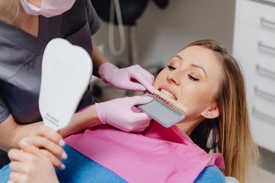
For crowns in the front of the mouth, feldspathic porcelain, a predominantly glass material with a non-crystalline structure, was for a time used primarily. A ceramicist began by making a plaster model from the dentist’s impression and adapting platinum foil around the resulting replica of the tooth preparation. Then, porcelain powder was added with a small, wet brush, building up the shape of the crown until it was ready to place into a furnace. The process was done multiple times to account for shrinkage of the material. Color was added during the process, matched to the original tooth using a shade guide that the dentist sent along with the impression and bite registration.
It’s still considered one of the most difficult accomplishments in restorative dentistry to match the color and form of one front tooth with the adjacent one. Sometimes, the teeth to be matched can have imperfections, color variations, or even cracks. Near the gum line, teeth are thicker, and thus darker, compared with biting surfaces, which are much thinner and more translucent as they’re composed only of enamel—a tooth is not a single color throughout.
Porcelain is a glass-like material; as such, it can crack or shatter when stressed in certain ways, and it can be abrasive to opposing teeth if chipped, so feldspathic porcelain is typically used for aesthetics, at the front of the mouth. Back teeth are meant to do the hard work, so harder materials are needed there.
Tougher Stuff
In the past, posterior crowns were made of gold or gold alloys. Crowns of porcelain fused to metal (PFM) were introduced in the 1960s, combining the inherent strength of a base layer of cast gold with the aesthetics of porcelain on the surface. PFM crowns were a good fit for the anterior, “aesthetic zone” too, and remained the standard for most crowns until quite recently.
As with other indirect restorations, impressions of the prepared tooth or teeth are taken and sent to a laboratory. For the meantime, a temporary crown can be made by first taking an impression of the original situation, or using a preformed temporary, usually made of resin.
PFM crowns are not without problems, however. When the gum margin recedes, a dark area of alloy material is visible around a crown’s base. People with nickel allergies must be wary of cheaper gold alloys. Additionally, different types of metals or fillings in the mouth can cause pain for some people, due to galvanic currents—electric charges generated by direct or indirect opposing restorations of differing conductivity.
A bridge—a replacement for a missing tooth created by crowning the adjacent teeth—cannot be made from porcelain alone, because it’s too weak. PFM enabled casting a single framework in a laboratory such that the three units would be joined together with porcelain fused to the outside, making a convincing stand-in for an entirely absent tooth. Of course, we now have the option of inserting a single implant into that missing space, avoiding preparation of the adjacent teeth. There are advantages and disadvantages for both procedures. We can talk more about implants later.
Stainless steel crowns exist too, and they’re mostly used in pediatric dentistry. The Hall Technique has been successfully applied for many years, requiring minimum preparation to seal in decay on baby molars.
Zirconia
Zirconia is a type of ceramic with a strong crystalline structure. Its original, tetragonal form is highly opaque with a flexural strength of about 1,100 megapascals (porcelain only reaches about 100 megapascals). Because of the high strength, crowns can be thinner and teeth can be prepared by removing only 0.6 to 1 millimeter from all surfaces; however, while metal can bend when enough force is applied, ceramic would instead crack. Its opacity relegates tetragonal zirconia to use farther back in the mouth, where strength considerations outweigh aesthetics.
Cubic zirconia (artificial diamond) can be added in different concentrations to achieve greater translucency and realism at the cost of strength. With 50 percent cubic zirconia, the appearance is much more suitable for anterior teeth, but flexural strength drops to about 600 megapascals.
Much like with PFM crowns, better color matching can be achieved by applying a surface layer of zirconia-reinforced lithium silicate. These surface layers tend to chip, reducing longevity, but the problem can be mitigated with an additional protective layer applied to the biting (occlusal) surface. Additionally, time has seen more-aesthetic staining and glazing techniques developed for use in the aesthetic zone.
Cementing zirconia is more demanding than with gold alloys. A crown must have its inner (intaglio) surface area increased at a laboratory by sandblasting with aluminum oxide particles. At a dental office, the finished crown is tried on, then washed with a special cleanser (salivary phospholipids would otherwise occupy many of the resin’s bonding sites). A ceramic primer with functional monomers bonds to teeth and zirconia, halting deterioration of collagen fibers. Dual-cure resin is used because zirconium’s opacity might prevent a curing light from doing the job.
The thickness of bonding and cementing agents needs to be such that it doesn’t interfere with the seating of any new crown or bridge. Bioactive cements can be used instead, but resin bonds with zirconia and lithium disilicate are much stronger.
Lithium Disilicate
Lithium disilicate is an aesthetic, high-strength polycrystalline (glass) ceramic first introduced in the 1990s. Unlike its predecessors, feldspathic and leucite-reinforced porcelains, lithium disilicate can be either conventionally cemented or adhesively bonded. Its strength is only 450 megapascals, so it’s not used farther back than the premolar areas, but it’s the standard choice for many dentists when crowning anterior teeth.
Biting surfaces need to be reduced by at least 1.5 millimeters in preparation for these crowns, and the material isn’t used for conventional bridges; however, it can work for Maryland bridges—a type of restoration, bonded to the back surface of one anterior tooth, with an “arm” that stretches out as an artificial tooth (pontic).
Although lithium disilicate can’t be adjusted before cementation, it can be repaired when chipped. Low concentrations of hydrofluoric acid etch or dissolve surface material, and silane coupling agents promote adhesion of replacement silica-based material. Hydrofluoric acid, rather than sandblasting, is used as well in laboratories when preparing the interior surfaces of new crowns or veneers.
CAD/CAM Technology
Over time, the predominant impression material has changed from polyether to polyvinyl siloxane; today, the option to use no material at all has become increasingly accessible. Conveniently, digital scanners can be used inside the mouth (intraorally). Entire sets of upper and lower teeth can be captured and sent directly to dental laboratories.
CAD/CAM (computer-aided design and computer-aided manufacturing) technologies are becoming more popular in dental offices all the time. The 3D models resulting from an intraoral scan can be mechanically milled into existence from ceramic blocks—right in the office! From tooth preparation to fitting a final crown, it can all be done in a single visit.
Smile design software coupled with CAD/CAM technology allows dentists and patients alike to preview and select from a range of shapes and sizes of teeth when considering how to restore a blemished smile. This is particularly useful when addressing parafunction or misalignment as well.
Dental advancements and the newer materials we’ve discussed have allowed the fabrication of lifelike crowns and bridges that are bonded to teeth rather than just cemented. Next time, we’ll explore veneers.